Q9UM54
Gene name |
MYO6 (KIAA0389) |
Protein name |
Unconventional myosin-VI |
Names |
Unconventional myosin-6 |
Species |
Homo sapiens (Human) |
KEGG Pathway |
hsa:4646 |
EC number |
|
Protein Class |
MYOSIN (PTHR13140) |
Descriptions
Myosin VI (Myo6) is the only minus-end directed nanomotor on actin and is essential for the structural integrity of the Golgi apparatus via the p53-dependent pro-survival pathway. The N-terminal region of the SAH domain folds back on the 3HB domain, thus adopting Myo6 to an elongated conformation that inhibits its activity. Notably, not all partners are capable of relieving the autoinhibition of Myo6. TOM1 and Dab2 are unable to bind the off-state of Myo6, while GIPC1 binds Myo6, releases its autoinhibition, and triggers proximal dimerization. The L926Q deafness mutation also disrupts the autoinhibition of Myo6 and indirectly impairs its proximal dimerization.
Autoinhibitory domains (AIDs)
Target domain |
834-917 (3HB domain) |
Relief mechanism |
Partner binding, Others |
Assay |
Structural analysis, Mutagenesis experiment |
Accessory elements
No accessory elements
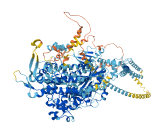
Autoinhibited structure
Activated structure

9 structures for Q9UM54
| Entry ID | Method | Resolution | Chain | Position | Source |
|---|---|---|---|---|---|
| 2N0Z | NMR | - | A | 1080-1122 | PDB |
| 2N10 | NMR | - | A | 1080-1131 | PDB |
| 2N11 | NMR | - | A | 998-1071 | PDB |
| 2N12 | NMR | - | A | 1050-1131 | PDB |
| 2N13 | NMR | - | A/D | 1080-1122 | PDB |
| 6E5N | NMR | - | B | 1050-1131 | PDB |
| 6J56 | X-ray | 180 A | A/B | 1166-1294 | PDB |
| 8W41 | EM | 354 A | A | 1-1294 | PDB |
| AF-Q9UM54-F1 | Predicted | AlphaFoldDB |
3 variants for Q9UM54
| Variant ID(s) | Position | Change | Description | Diseaes Association | Provenance |
|---|---|---|---|---|---|
|
VAR_016209 rs121912559 |
216 | E>V | DFNB37 [UniProt] | Yes |
UniProt dbSNP |
|
rs121912560 VAR_029988 |
246 | H>R | DFNHCM [UniProt] | Yes |
UniProt dbSNP |
| VAR_012110 | 442 | C>Y | DFNA22 [UniProt] | Yes | UniProt |
3 associated diseases with Q9UM54
[MIM: 606346]: Deafness, autosomal dominant, 22 (DFNA22)
A form of non-syndromic sensorineural hearing loss. Sensorineural deafness results from damage to the neural receptors of the inner ear, the nerve pathways to the brain, or the area of the brain that receives sound information. DFNA22 is progressive and postlingual, with onset during childhood. By the age of approximately 50 years, affected individuals invariably have profound sensorineural deafness. . Note=The disease is caused by variants affecting the gene represented in this entry.
[MIM: 607821]: Deafness, autosomal recessive, 37 (DFNB37)
A form of non-syndromic sensorineural hearing loss. Sensorineural deafness results from damage to the neural receptors of the inner ear, the nerve pathways to the brain, or the area of the brain that receives sound information. . Note=The disease is caused by variants affecting the gene represented in this entry.
[MIM: 606346]: Deafness, autosomal dominant 22, with hypertrophic cardiomyopathy (DFNHCM)
An autosomal dominant sensorineural deafness associated with hypertrophic cardiomyopathy. . Note=The disease is caused by variants affecting the gene represented in this entry.
Without disease ID
- A form of non-syndromic sensorineural hearing loss. Sensorineural deafness results from damage to the neural receptors of the inner ear, the nerve pathways to the brain, or the area of the brain that receives sound information. DFNA22 is progressive and postlingual, with onset during childhood. By the age of approximately 50 years, affected individuals invariably have profound sensorineural deafness. . Note=The disease is caused by variants affecting the gene represented in this entry.
- A form of non-syndromic sensorineural hearing loss. Sensorineural deafness results from damage to the neural receptors of the inner ear, the nerve pathways to the brain, or the area of the brain that receives sound information. . Note=The disease is caused by variants affecting the gene represented in this entry.
- An autosomal dominant sensorineural deafness associated with hypertrophic cardiomyopathy. . Note=The disease is caused by variants affecting the gene represented in this entry.
5 regional properties for Q9UM54
| Type | Name | Position | InterPro Accession |
|---|---|---|---|
| domain | Myosin head, motor domain | 51 - 772 | IPR001609 |
| domain | Myosin, N-terminal, SH3-like | 2 - 53 | IPR004009 |
| domain | Myosin VI, cargo binding domain | 1177 - 1267 | IPR032412 |
| domain | Class VI myosin, motor domain | 71 - 759 | IPR036114 |
| domain | Myosin VI, lever arm | 770 - 917 | IPR049016 |
Functions
| Description | ||
|---|---|---|
| EC Number | ||
| Subcellular Localization |
|
|
| PANTHER Family | PTHR13140 | MYOSIN |
| PANTHER Subfamily | PTHR13140:SF745 | UNCONVENTIONAL MYOSIN-VI |
| PANTHER Protein Class | actin binding motor protein | |
| PANTHER Pathway Category |
Nicotinic acetylcholine receptor signaling pathway Myosin |
|
27 GO annotations of cellular component
| Name | Definition |
|---|---|
| actin cytoskeleton | The part of the cytoskeleton (the internal framework of a cell) composed of actin and associated proteins. Includes actin cytoskeleton-associated complexes. |
| actin filament | A filamentous structure formed of a two-stranded helical polymer of the protein actin and associated proteins. Actin filaments are a major component of the contractile apparatus of skeletal muscle and the microfilaments of the cytoskeleton of eukaryotic cells. The filaments, comprising polymerized globular actin molecules, appear as flexible structures with a diameter of 5-9 nm. They are organized into a variety of linear bundles, two-dimensional networks, and three dimensional gels. In the cytoskeleton they are most highly concentrated in the cortex of the cell just beneath the plasma membrane. |
| apical part of cell | The region of a polarized cell that forms a tip or is distal to a base. For example, in a polarized epithelial cell, the apical region has an exposed surface and lies opposite to the basal lamina that separates the epithelium from other tissue. |
| autophagosome | A double-membrane-bounded compartment that engulfs endogenous cellular material as well as invading microorganisms to target them to the lytic vacuole/lysosome for degradation as part of macroautophagy. |
| cell cortex | The region of a cell that lies just beneath the plasma membrane and often, but not always, contains a network of actin filaments and associated proteins. |
| clathrin-coated pit | A part of the endomembrane system in the form of an invagination of a membrane upon which a clathrin coat forms, and that can be converted by vesicle budding into a clathrin-coated vesicle. Coated pits form on the plasma membrane, where they are involved in receptor-mediated selective transport of many proteins and other macromolecules across the cell membrane, in the trans-Golgi network, and on some endosomes. |
| clathrin-coated vesicle membrane | The lipid bilayer surrounding a clathrin-coated vesicle. |
| cytoplasm | The contents of a cell excluding the plasma membrane and nucleus, but including other subcellular structures. |
| cytoplasmic vesicle | A vesicle found in the cytoplasm of a cell. |
| cytosol | The part of the cytoplasm that does not contain organelles but which does contain other particulate matter, such as protein complexes. |
| endocytic vesicle | A membrane-bounded intracellular vesicle formed by invagination of the plasma membrane around an extracellular substance. Endocytic vesicles fuse with early endosomes to deliver the cargo for further sorting. |
| endosome | A vacuole to which materials ingested by endocytosis are delivered. |
| extracellular exosome | A vesicle that is released into the extracellular region by fusion of the limiting endosomal membrane of a multivesicular body with the plasma membrane. Extracellular exosomes, also simply called exosomes, have a diameter of about 40-100 nm. |
| filamentous actin | A two-stranded helical polymer of the protein actin. |
| filopodium | Thin, stiff, actin-based protrusion extended by the leading edge of a motile cell such as a crawling fibroblast or amoeba, or an axonal or dendritic growth cone, or a dendritic shaft. |
| Golgi apparatus | A membrane-bound cytoplasmic organelle of the endomembrane system that further processes the core oligosaccharides (e.g. N-glycans) added to proteins in the endoplasmic reticulum and packages them into membrane-bound vesicles. The Golgi apparatus operates at the intersection of the secretory, lysosomal, and endocytic pathways. |
| lysosomal membrane | The lipid bilayer surrounding the lysosome and separating its contents from the cell cytoplasm. |
| membrane | A lipid bilayer along with all the proteins and protein complexes embedded in it and attached to it. |
| microvillus | Thin cylindrical membrane-covered projections on the surface of an animal cell containing a core bundle of actin filaments. Present in especially large numbers on the absorptive surface of intestinal cells. |
| nuclear membrane | Either of the lipid bilayers that surround the nucleus and form the nuclear envelope; excludes the intermembrane space. |
| nucleoplasm | That part of the nuclear content other than the chromosomes or the nucleolus. |
| nucleus | A membrane-bounded organelle of eukaryotic cells in which chromosomes are housed and replicated. In most cells, the nucleus contains all of the cell's chromosomes except the organellar chromosomes, and is the site of RNA synthesis and processing. In some species, or in specialized cell types, RNA metabolism or DNA replication may be absent. |
| perinuclear region of cytoplasm | Cytoplasm situated near, or occurring around, the nucleus. |
| plasma membrane | The membrane surrounding a cell that separates the cell from its external environment. It consists of a phospholipid bilayer and associated proteins. |
| ruffle | Projection at the leading edge of a crawling cell; the protrusions are supported by a microfilament meshwork. |
| ruffle membrane | The portion of the plasma membrane surrounding a ruffle. |
| unconventional myosin complex | A portmanteau term for myosins other than myosin II. |
9 GO annotations of molecular function
| Name | Definition |
|---|---|
| actin binding | Binding to monomeric or multimeric forms of actin, including actin filaments. |
| actin filament binding | Binding to an actin filament, also known as F-actin, a helical filamentous polymer of globular G-actin subunits. |
| ADP binding | Binding to ADP, adenosine 5'-diphosphate. |
| ATP binding | Binding to ATP, adenosine 5'-triphosphate, a universally important coenzyme and enzyme regulator. |
| calmodulin binding | Binding to calmodulin, a calcium-binding protein with many roles, both in the calcium-bound and calcium-free states. |
| cytoskeletal motor activity | Generation of force resulting in movement, for example along a microfilament or microtubule, or in torque resulting in membrane scission or rotation of a flagellum. The energy required is obtained either from the hydrolysis of a nucleoside triphosphate or by an electrochemical proton gradient (proton-motive force). |
| identical protein binding | Binding to an identical protein or proteins. |
| microfilament motor activity | A motor activity that generates movement along a microfilament, driven by ATP hydrolysis. |
| minus-end directed microfilament motor activity | A motor activity that generates movement along a microfilament towards the minus end, driven by ATP hydrolysis. The minus end of an actin filament is the end that does not preferentially add actin monomers. |
11 GO annotations of biological process
| Name | Definition |
|---|---|
| actin filament organization | A process that is carried out at the cellular level which results in the assembly, arrangement of constituent parts, or disassembly of cytoskeletal structures comprising actin filaments. Includes processes that control the spatial distribution of actin filaments, such as organizing filaments into meshworks, bundles, or other structures, as by cross-linking. |
| actin filament-based movement | Movement of organelles or other particles along actin filaments, or sliding of actin filaments past each other, mediated by motor proteins. |
| DNA damage response, signal transduction by p53 class mediator | A cascade of processes induced by the cell cycle regulator phosphoprotein p53, or an equivalent protein, in response to the detection of DNA damage. |
| endocytosis | A vesicle-mediated transport process in which cells take up external materials or membrane constituents by the invagination of a part of the plasma membrane to form a new membrane-bounded vesicle. |
| inner ear auditory receptor cell differentiation | The process in which a relatively unspecialized inner cell acquires specialized features of an auditory hair cell. |
| inner ear morphogenesis | The process in which the anatomical structures of the inner ear are generated and organized. The inner ear is the structure in vertebrates that contains the organs of balance and hearing. It consists of soft hollow sensory structures (the membranous labyrinth) containing fluid (endolymph) surrounded by fluid (perilymph) and encased in a bony cavity (the bony labyrinth). It consists of two chambers, the sacculus and utriculus, from which arise the cochlea and semicircular canals respectively. |
| intracellular protein transport | The directed movement of proteins in a cell, including the movement of proteins between specific compartments or structures within a cell, such as organelles of a eukaryotic cell. |
| regulation of secretion | Any process that modulates the frequency, rate or extent of the controlled release of a substance from a cell or a tissue. |
| response to xenobiotic stimulus | Any process that results in a change in state or activity of a cell or an organism (in terms of movement, secretion, enzyme production, gene expression, etc.) as a result of a stimulus from a xenobiotic, a compound foreign to the organim exposed to it. It may be synthesized by another organism (like ampicilin) or it can be a synthetic chemical. |
| sensory perception of sound | The series of events required for an organism to receive an auditory stimulus, convert it to a molecular signal, and recognize and characterize the signal. Sonic stimuli are detected in the form of vibrations and are processed to form a sound. |
| vesicle transport along actin filament | Movement of a vesicle along an actin filament, mediated by motor proteins. |
11 homologous proteins in AiPD
| UniProt AC | Gene Name | Protein Name | Species | Evidence Code |
|---|---|---|---|---|
| E1BPK6 | MYO6 | Unconventional myosin-VI | Bos taurus (Bovine) | SS |
| Q9I8D1 | MYO6 | Unconventional myosin-VI | Gallus gallus (Chicken) | SS |
| B0I1T2 | MYO1G | Unconventional myosin-Ig | Homo sapiens (Human) | PR |
| Q12965 | MYO1E | Unconventional myosin-Ie | Homo sapiens (Human) | PR |
| O00160 | MYO1F | Unconventional myosin-If | Homo sapiens (Human) | PR |
| Q9Y4I1 | MYO5A | Unconventional myosin-Va | Homo sapiens (Human) | SS |
| Q9NQX4 | MYO5C | Unconventional myosin-Vc | Homo sapiens (Human) | SS |
| Q9ULV0 | MYO5B | Unconventional myosin-Vb | Homo sapiens (Human) | SS |
| Q64331 | Myo6 | Unconventional myosin-VI | Mus musculus (Mouse) | SS |
| Q29122 | MYO6 | Unconventional myosin-VI | Sus scrofa (Pig) | SS |
| Q9M2K0 | XI-J | Myosin-16 | Arabidopsis thaliana (Mouse-ear cress) | PR |
| 10 | 20 | 30 | 40 | 50 | 60 |
| MEDGKPVWAP | HPTDGFQMGN | IVDIGPDSLT | IEPLNQKGKT | FLALINQVFP | AEEDSKKDVE |
| 70 | 80 | 90 | 100 | 110 | 120 |
| DNCSLMYLNE | ATLLHNIKVR | YSKDRIYTYV | ANILIAVNPY | FDIPKIYSSE | AIKSYQGKSL |
| 130 | 140 | 150 | 160 | 170 | 180 |
| GTRPPHVFAI | ADKAFRDMKV | LKMSQSIIVS | GESGAGKTEN | TKFVLRYLTE | SYGTGQDIDD |
| 190 | 200 | 210 | 220 | 230 | 240 |
| RIVEANPLLE | AFGNAKTVRN | NNSSRFGKFV | EIHFNEKSSV | VGGFVSHYLL | EKSRICVQGK |
| 250 | 260 | 270 | 280 | 290 | 300 |
| EERNYHIFYR | LCAGASEDIR | EKLHLSSPDN | FRYLNRGCTR | YFANKETDKQ | ILQNRKSPEY |
| 310 | 320 | 330 | 340 | 350 | 360 |
| LKAGSMKDPL | LDDHGDFIRM | CTAMKKIGLD | DEEKLDLFRV | VAGVLHLGNI | DFEEAGSTSG |
| 370 | 380 | 390 | 400 | 410 | 420 |
| GCNLKNKSAQ | SLEYCAELLG | LDQDDLRVSL | TTRVMLTTAG | GTKGTVIKVP | LKVEQANNAR |
| 430 | 440 | 450 | 460 | 470 | 480 |
| DALAKTVYSH | LFDHVVNRVN | QCFPFETSSY | FIGVLDIAGF | EYFEHNSFEQ | FCINYCNEKL |
| 490 | 500 | 510 | 520 | 530 | 540 |
| QQFFNERILK | EEQELYQKEG | LGVNEVHYVD | NQDCIDLIEA | KLVGILDILD | EENRLPQPSD |
| 550 | 560 | 570 | 580 | 590 | 600 |
| QHFTSAVHQK | HKDHFRLTIP | RKSKLAVHRN | IRDDEGFIIR | HFAGAVCYET | TQFVEKNNDA |
| 610 | 620 | 630 | 640 | 650 | 660 |
| LHMSLESLIC | ESRDKFIREL | FESSTNNNKD | TKQKAGKLSF | ISVGNKFKTQ | LNLLLDKLRS |
| 670 | 680 | 690 | 700 | 710 | 720 |
| TGASFIRCIK | PNLKMTSHHF | EGAQILSQLQ | CSGMVSVLDL | MQGGYPSRAS | FHELYNMYKK |
| 730 | 740 | 750 | 760 | 770 | 780 |
| YMPDKLARLD | PRLFCKALFK | ALGLNENDYK | FGLTKVFFRP | GKFAEFDQIM | KSDPDHLAEL |
| 790 | 800 | 810 | 820 | 830 | 840 |
| VKRVNHWLTC | SRWKKVQWCS | LSVIKLKNKI | KYRAEACIKM | QKTIRMWLCK | RRHKPRIDGL |
| 850 | 860 | 870 | 880 | 890 | 900 |
| VKVGTLKKRL | DKFNEVVSVL | KDGKPEMNKQ | IKNLEISIDT | LMAKIKSTMM | TQEQIQKEYD |
| 910 | 920 | 930 | 940 | 950 | 960 |
| ALVKSSEELL | SALQKKKQQE | EEAERLRRIQ | EEMEKERKRR | EEDEKRRRKE | EEERRMKLEM |
| 970 | 980 | 990 | 1000 | 1010 | 1020 |
| EAKRKQEEEE | RKKREDDEKR | IQAEVEAQLA | RQKEEESQQQ | AVLEQERRDR | ELALRIAQSE |
| 1030 | 1040 | 1050 | 1060 | 1070 | 1080 |
| AELISDEAQA | DLALRRSLDS | YPVSKNDGTR | PKMTPEQMAK | EMSEFLSRGP | AVLATKAAAG |
| 1090 | 1100 | 1110 | 1120 | 1130 | 1140 |
| TKKYDLSKWK | YAELRDTINT | SCDIELLAAC | REEFHRRLKV | YHAWKSKNKK | RNTETEQRAP |
| 1150 | 1160 | 1170 | 1180 | 1190 | 1200 |
| KSVTDYDFAP | FLNNSPQQNP | AAQIPARQRE | IEMNRQQRFF | RIPFIRPADQ | YKDPQSKKKG |
| 1210 | 1220 | 1230 | 1240 | 1250 | 1260 |
| WWYAHFDGPW | IARQMELHPD | KPPILLVAGK | DDMEMCELNL | EETGLTRKRG | AEILPRQFEE |
| 1270 | 1280 | 1290 | |||
| IWERCGGIQY | LQNAIESRQA | RPTYATAMLQ | SLLK |