Descriptions
The autoinhibited protein was predicted that may have potential autoinhibitory elements via cis-regPred.
Autoinhibitory domains (AIDs)
Target domain |
|
Relief mechanism |
|
Assay |
cis-regPred |
Accessory elements
No accessory elements
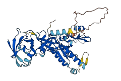
Autoinhibited structure

Activated structure
1 structures for Q9QYH1
| Entry ID | Method | Resolution | Chain | Position | Source |
|---|---|---|---|---|---|
| AF-Q9QYH1-F1 | Predicted | AlphaFoldDB |
No variants for Q9QYH1
| Variant ID(s) | Position | Change | Description | Diseaes Association | Provenance |
|---|---|---|---|---|---|
| No variants for Q9QYH1 | |||||
No associated diseases with Q9QYH1
6 regional properties for Q9QYH1
| Type | Name | Position | InterPro Accession |
|---|---|---|---|
| domain | SH3 domain | 91 - 161 | IPR001452 |
| domain | PDZ domain | 3 - 84 | IPR001478 |
| domain | Guanylate kinase-like domain | 232 - 421 | IPR008144 |
| domain | Guanylate kinase/L-type calcium channel beta subunit | 231 - 424 | IPR008145 |
| conserved_site | Guanylate kinase, conserved site | 267 - 284 | IPR020590 |
| domain | MPP4, SH3 domain | 95 - 155 | IPR035600 |
10 GO annotations of cellular component
| Name | Definition |
|---|---|
| adherens junction | A cell-cell junction composed of the epithelial cadherin-catenin complex. The epithelial cadherins, or E-cadherins, of each interacting cell extend through the plasma membrane into the extracellular space and bind to each other. The E-cadherins bind to catenins on the cytoplasmic side of the membrane, where the E-cadherin-catenin complex binds to cytoskeletal components and regulatory and signaling molecules. |
| basal part of cell | The region of a cell situated near the base. For example, in a polarized epithelial cell, the basal surface rests on the basal lamina that separates the epithelium from other tissue. |
| cell-cell junction | A cell junction that forms a connection between two or more cells of an organism; excludes direct cytoplasmic intercellular bridges, such as ring canals in insects. |
| cone cell pedicle | A specialized axon terminus which is produced by retinal cone cells. Pedicles are large, conical, flat end-feet (8-10 micrometers diameter) of the retinal cone axon that lie more or less side by side on the same plane at the outer edge of the outer plexiform layer (OPL). |
| Golgi membrane | The lipid bilayer surrounding any of the compartments of the Golgi apparatus. |
| lateral plasma membrane | The portion of the plasma membrane at the lateral side of the cell. In epithelial cells, lateral plasma membranes are on the sides of cells which lie at the interface of adjacent cells. |
| plasma membrane | The membrane surrounding a cell that separates the cell from its external environment. It consists of a phospholipid bilayer and associated proteins. |
| presynaptic membrane | A specialized area of membrane of the axon terminal that faces the plasma membrane of the neuron or muscle fiber with which the axon terminal establishes a synaptic junction; many synaptic junctions exhibit structural presynaptic characteristics, such as conical, electron-dense internal protrusions, that distinguish it from the remainder of the axon plasma membrane. |
| protein-containing complex | A stable assembly of two or more macromolecules, i.e. proteins, nucleic acids, carbohydrates or lipids, in which at least one component is a protein and the constituent parts function together. |
| rod spherule | A specialized neuron projection which is the site of synaptic transmission produced by retinal rod cells. Rod spherules are small round enlargements of the axon (3-5 micrometers diameter) or even extensions of the cell body. |
1 GO annotations of molecular function
| Name | Definition |
|---|---|
| signaling receptor binding | Binding to one or more specific sites on a receptor molecule, a macromolecule that undergoes combination with a hormone, neurotransmitter, drug or intracellular messenger to initiate a change in cell function. |
1 GO annotations of biological process
| Name | Definition |
|---|---|
| protein localization to synapse | Any process in which a protein is transported to, and/or maintained at the synapse, the junction between a nerve fiber of one neuron and another neuron or muscle fiber or glial cell. |
14 homologous proteins in AiPD
| UniProt AC | Gene Name | Protein Name | Species | Evidence Code |
|---|---|---|---|---|
| Q24210 | CASK | Peripheral plasma membrane protein CASK | Drosophila melanogaster (Fruit fly) | SS |
| Q9NZW5 | PALS2 | Protein PALS2 | Homo sapiens (Human) | PR |
| Q00013 | MPP1 | 55 kDa erythrocyte membrane protein | Homo sapiens (Human) | PR |
| O14936 | CASK | Peripheral plasma membrane protein CASK | Homo sapiens (Human) | EV |
| Q14168 | MPP2 | MAGUK p55 subfamily member 2 | Homo sapiens (Human) | PR |
| Q96JB8 | MPP4 | MAGUK p55 subfamily member 4 | Homo sapiens (Human) | PR |
| Q9WV34 | Mpp2 | MAGUK p55 subfamily member 2 | Mus musculus (Mouse) | PR |
| O70589 | Cask | Peripheral plasma membrane protein CASK | Mus musculus (Mouse) | SS |
| P70290 | Mpp1 | 55 kDa erythrocyte membrane protein | Mus musculus (Mouse) | PR |
| O88910 | Mpp3 | MAGUK p55 subfamily member 3 | Mus musculus (Mouse) | PR |
| Q8BVD5 | Mpp7 | MAGUK p55 subfamily member 7 | Mus musculus (Mouse) | PR |
| Q9JLB0 | Pals2 | Protein PALS2 | Mus musculus (Mouse) | PR |
| Q6P7F1 | Mpp4 | MAGUK p55 subfamily member 4 | Mus musculus (Mouse) | PR |
| P54936 | lin-2 | Protein lin-2 | Caenorhabditis elegans | SS |
| 10 | 20 | 30 | 40 | 50 | 60 |
| MRTVCLVKNQ | QPLGATIKRH | EITGDILVAR | VIHGGLVERN | GLLYAGDKLV | EVNGVPVEGL |
| 70 | 80 | 90 | 100 | 110 | 120 |
| DPEQVIHILA | MSCGTIMFKV | IPVSAPPVSS | QTTVYVRAMI | DYWPQEDPDI | PCMDAGLPFL |
| 130 | 140 | 150 | 160 | 170 | 180 |
| KGDILQIVDQ | SDALWWQARK | ISDIAICAGL | IPSNHLLKRK | QREFWWSQPY | QPHTCLKSTR |
| 190 | 200 | 210 | 220 | 230 | 240 |
| SKEEFVGDGQ | QFFIAGFRQQ | HANMRCTCSC | YSAVGAPYEE | VVRYQRQPAD | KHRLIVLVGP |
| 250 | 260 | 270 | 280 | 290 | 300 |
| SGVGVNELRR | QLIGCNPSCF | QSAVPHTTRS | PKSYEMDGRE | YHYVSRETFE | SLMYGHRMLE |
| 310 | 320 | 330 | 340 | 350 | 360 |
| FGEYKGHLYG | TSVNAVLAVL | DEGKICVMDL | EPQDIQLART | RELKPYVIFI | KPPSMSSMRH |
| 370 | 380 | 390 | 400 | 410 | 420 |
| SRRNAKIITD | YFVDMKFKDE | DLQEMEELAQ | KMESQFGQFF | DHVIVNDNLQ | DARAQLLSAI |
| 430 | 440 | ||||
| QKAEEELQWV | PEAWVSPGAE | S |