Q9JLT0
Gene name |
Myh10 |
Protein name |
Myosin-10 |
Names |
Cellular myosin heavy chain, type B , Myosin heavy chain 10 , Myosin heavy chain, non-muscle IIb , Non-muscle myosin heavy chain B , NMMHC-B , Non-muscle myosin heavy chain IIb , NMMHC II-b , NMMHC-IIB |
Species |
Rattus norvegicus (Rat) |
KEGG Pathway |
rno:79433 |
EC number |
|
Protein Class |
|
Descriptions
Autoinhibitory domains (AIDs)
Target domain |
79-784 (Myosin head, motor domain) |
Relief mechanism |
Partner binding |
Assay |
|
Accessory elements
No accessory elements
References
- Yeon JH et al. (2016) "Systems-wide Identification of cis-Regulatory Elements in Proteins", Cell systems, 2, 89-100
- Morck MM et al. (2022) "Hypertrophic cardiomyopathy mutations in the pliant and light chain-binding regions of the lever arm of human β-cardiac myosin have divergent effects on myosin function", eLife, 11,
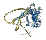
Autoinhibited structure

Activated structure
1 structures for Q9JLT0
| Entry ID | Method | Resolution | Chain | Position | Source |
|---|---|---|---|---|---|
| AF-Q9JLT0-F1 | Predicted | AlphaFoldDB |
No variants for Q9JLT0
| Variant ID(s) | Position | Change | Description | Diseaes Association | Provenance |
|---|---|---|---|---|---|
| No variants for Q9JLT0 | |||||
No associated diseases with Q9JLT0
24 GO annotations of cellular component
| Name | Definition |
|---|---|
| actomyosin | Any complex of actin, myosin, and accessory proteins. |
| axon | The long process of a neuron that conducts nerve impulses, usually away from the cell body to the terminals and varicosities, which are sites of storage and release of neurotransmitter. |
| brush border | The dense covering of microvilli on the apical surface of an epithelial cell in tissues such as the intestine, kidney, and choroid plexus; the microvilli aid absorption by increasing the surface area of the cell. |
| cell cortex | The region of a cell that lies just beneath the plasma membrane and often, but not always, contains a network of actin filaments and associated proteins. |
| cleavage furrow | The cleavage furrow is a plasma membrane invagination at the cell division site. The cleavage furrow begins as a shallow groove and eventually deepens to divide the cytoplasm. |
| cytoplasm | The contents of a cell excluding the plasma membrane and nucleus, but including other subcellular structures. |
| cytoplasmic side of plasma membrane | The leaflet the plasma membrane that faces the cytoplasm and any proteins embedded or anchored in it or attached to its surface. |
| cytosol | The part of the cytoplasm that does not contain organelles but which does contain other particulate matter, such as protein complexes. |
| dendritic spine | A small, membranous protrusion from a dendrite that forms a postsynaptic compartment, typically receiving input from a single presynapse. They function as partially isolated biochemical and an electrical compartments. Spine morphology is variable:they can be thin, stubby, mushroom, or branched, with a continuum of intermediate morphologies. They typically terminate in a bulb shape, linked to the dendritic shaft by a restriction. Spine remodeling is though to be involved in synaptic plasticity. |
| glutamatergic synapse | A synapse that uses glutamate as a neurotransmitter. |
| growth cone | The migrating motile tip of a growing neuron projection, where actin accumulates, and the actin cytoskeleton is the most dynamic. |
| lamellipodium | A thin sheetlike process extended by the leading edge of a migrating cell or extending cell process; contains a dense meshwork of actin filaments. |
| midbody | A thin cytoplasmic bridge formed between daughter cells at the end of cytokinesis. The midbody forms where the contractile ring constricts, and may persist for some time before finally breaking to complete cytokinesis. |
| myosin complex | A protein complex, formed of one or more myosin heavy chains plus associated light chains and other proteins, that functions as a molecular motor; uses the energy of ATP hydrolysis to move actin filaments or to move vesicles or other cargo on fixed actin filaments; has magnesium-ATPase activity and binds actin. Myosin classes are distinguished based on sequence features of the motor, or head, domain, but also have distinct tail regions that are believed to bind specific cargoes. |
| myosin filament | A supramolecular fiber containing myosin heavy chains, plus associated light chains and other proteins, in which the myosin heavy chains are arranged into a filament. |
| myosin II complex | A myosin complex containing two class II myosin heavy chains, two myosin essential light chains and two myosin regulatory light chains. Also known as classical myosin or conventional myosin, the myosin II class includes the major muscle myosin of vertebrate and invertebrate muscle, and is characterized by alpha-helical coiled coil tails that self assemble to form a variety of filament structures. |
| myosin II filament | A bipolar filament composed of myosin II molecules. |
| neuromuscular junction | The junction between the axon of a motor neuron and a muscle fiber. In response to the arrival of action potentials, the presynaptic button releases molecules of neurotransmitters into the synaptic cleft. These diffuse across the cleft and transmit the signal to the postsynaptic membrane of the muscle fiber, leading to a change in post-synaptic potential. |
| neuron projection | A prolongation or process extending from a nerve cell, e.g. an axon or dendrite. |
| neuronal cell body | The portion of a neuron that includes the nucleus, but excludes cell projections such as axons and dendrites. |
| postsynaptic actin cytoskeleton | The actin cytoskeleton that is part of a postsynapse. |
| sarcoplasm | The cytoplasm of a muscle cell; includes the sarcoplasmic reticulum. |
| spindle | The array of microtubules and associated molecules that forms between opposite poles of a eukaryotic cell during mitosis or meiosis and serves to move the duplicated chromosomes apart. |
| stress fiber | A contractile actin filament bundle that consists of short actin filaments with alternating polarity, cross-linked by alpha-actinin and possibly other actin bundling proteins, and with myosin present in a periodic distribution along the fiber. |
8 GO annotations of molecular function
| Name | Definition |
|---|---|
| actin filament binding | Binding to an actin filament, also known as F-actin, a helical filamentous polymer of globular G-actin subunits. |
| ADP binding | Binding to ADP, adenosine 5'-diphosphate. |
| ATP binding | Binding to ATP, adenosine 5'-triphosphate, a universally important coenzyme and enzyme regulator. |
| calmodulin binding | Binding to calmodulin, a calcium-binding protein with many roles, both in the calcium-bound and calcium-free states. |
| identical protein binding | Binding to an identical protein or proteins. |
| microfilament motor activity | A motor activity that generates movement along a microfilament, driven by ATP hydrolysis. |
| mRNA 5'-UTR binding | Binding to an mRNA molecule at its 5' untranslated region. |
| RNA stem-loop binding | Binding to a stem-loop in an RNA molecule. An RNA stem-loop is a secondary RNA structure consisting of a double-stranded RNA (dsRNA) stem and a terminal loop. |
36 GO annotations of biological process
| Name | Definition |
|---|---|
| actin cytoskeleton organization | A process that is carried out at the cellular level which results in the assembly, arrangement of constituent parts, or disassembly of cytoskeletal structures comprising actin filaments and their associated proteins. |
| actin filament bundle assembly | The assembly of actin filament bundles; actin filaments are on the same axis but may be oriented with the same or opposite polarities and may be packed with different levels of tightness. |
| actin filament bundle distribution | Any cellular process that establishes the spatial arrangement of actin filament bundles within the cell. |
| actin filament-based movement | Movement of organelles or other particles along actin filaments, or sliding of actin filaments past each other, mediated by motor proteins. |
| actomyosin structure organization | A process that is carried out at the cellular level which results in the assembly, arrangement of constituent parts, or disassembly of cytoskeletal structures containing both actin and myosin or paramyosin. The myosin may be organized into filaments. |
| adult heart development | The process whose specific outcome is the progression of the adult heart over time, from its formation to the mature structure. |
| aorta development | The progression of the aorta over time, from its initial formation to the mature structure. An aorta is an artery that carries blood from the heart to other parts of the body. |
| axon guidance | The chemotaxis process that directs the migration of an axon growth cone to a specific target site in response to a combination of attractive and repulsive cues. |
| axonogenesis | De novo generation of a long process of a neuron, including the terminal branched region. Refers to the morphogenesis or creation of shape or form of the developing axon, which carries efferent (outgoing) action potentials from the cell body towards target cells. |
| brain development | The process whose specific outcome is the progression of the brain over time, from its formation to the mature structure. Brain development begins with patterning events in the neural tube and ends with the mature structure that is the center of thought and emotion. The brain is responsible for the coordination and control of bodily activities and the interpretation of information from the senses (sight, hearing, smell, etc.). |
| cardiac muscle cell proliferation | The expansion of a cardiac muscle cell population by cell division. |
| cardiac myofibril assembly | The process whose specific outcome is the progression of the cardiac myofibril over time, from its formation to the mature structure. A cardiac myofibril is a myofibril specific to cardiac muscle cells. |
| cardiac septum development | The progression of a cardiac septum over time, from its initial formation to the mature structure. |
| cell adhesion | The attachment of a cell, either to another cell or to an underlying substrate such as the extracellular matrix, via cell adhesion molecules. |
| cerebellar Purkinje cell layer development | The process whose specific outcome is the progression of the cerebellar Purkinje cell layer over time, from its formation to the mature structure. The Purkinje cell layer lies just underneath the molecular layer of the cerebellar cortex. It contains the neuronal cell bodies of the Purkinje cells that are arranged side by side in a single layer. Candelabrum interneurons are vertically oriented between the Purkinje cells. Purkinje neurons are inhibitory and provide the output of the cerebellar cortex through axons that project into the white matter. Extensive dendritic trees from the Purkinje cells extend upward in a single plane into the molecular layer where they synapse with parallel fibers of granule cells. |
| coronary vasculature development | The process whose specific outcome is the progression of the blood vessels of the heart over time, from its formation to the mature structure. |
| exocytosis | A process of secretion by a cell that results in the release of intracellular molecules (e.g. hormones, matrix proteins) contained within a membrane-bounded vesicle. Exocytosis can occur either by full fusion, when the vesicle collapses into the plasma membrane, or by a kiss-and-run mechanism that involves the formation of a transient contact, a pore, between a granule (for exemple of chromaffin cells) and the plasma membrane. The latter process most of the time leads to only partial secretion of the granule content. Exocytosis begins with steps that prepare vesicles for fusion with the membrane (tethering and docking) and ends when molecules are secreted from the cell. |
| fourth ventricle development | The process whose specific outcome is the progression of the fourth ventricle over time, from its formation to the mature structure. The fourth ventricle is an irregularly shaped cavity in the rhombencephalon, between the medulla oblongata, the pons, and the isthmus in front, and the cerebellum behind. It is continuous with the central canal of the cord below and with the cerebral aqueduct above, and through its lateral and median apertures it communicates with the subarachnoid space. |
| heart development | The process whose specific outcome is the progression of the heart over time, from its formation to the mature structure. The heart is a hollow, muscular organ, which, by contracting rhythmically, keeps up the circulation of the blood. |
| in utero embryonic development | The process whose specific outcome is the progression of the embryo in the uterus over time, from formation of the zygote in the oviduct, to birth. An example of this process is found in Mus musculus. |
| lateral ventricle development | The process whose specific outcome is the progression of the lateral ventricles over time, from the formation to the mature structure. The two lateral ventricles are a cavity in each of the cerebral hemispheres derived from the cavity of the embryonic neural tube. They are separated from each other by the septum pellucidum, and each communicates with the third ventricle by the foramen of Monro, through which also the choroid plexuses of the lateral ventricles become continuous with that of the third ventricle. |
| mitotic cytokinesis | A cell cycle process that results in the division of the cytoplasm of a cell after mitosis, resulting in the separation of the original cell into two daughter cells. |
| myofibril assembly | Formation of myofibrils, the repeating units of striated muscle. |
| neuromuscular process controlling balance | Any process that an organism uses to control its balance, the orientation of the organism (or the head of the organism) in relation to the source of gravity. In humans and animals, balance is perceived through visual cues, the labyrinth system of the inner ears and information from skin pressure receptors and muscle and joint receptors. |
| neuron migration | The characteristic movement of an immature neuron from germinal zones to specific positions where they will reside as they mature. |
| neuron projection development | The process whose specific outcome is the progression of a neuron projection over time, from its formation to the mature structure. A neuron projection is any process extending from a neural cell, such as axons or dendrites (collectively called neurites). |
| nuclear migration | The directed movement of the nucleus to a specific location within a cell. |
| plasma membrane repair | The resealing of a cell plasma membrane after cellular wounding due to, for instance, mechanical stress. |
| positive regulation of protein secretion | Any process that activates or increases the frequency, rate or extent of the controlled release of a protein from a cell. |
| postsynaptic actin cytoskeleton organization | A process that is carried out at the cellular level which results in the assembly, arrangement of constituent parts, or disassembly of cytoskeletal structures comprising actin filaments and their associated proteins in the postsynaptic actin cytoskeleton. |
| regulation of cell shape | Any process that modulates the surface configuration of a cell. |
| regulation of modification of postsynaptic actin cytoskeleton | Any process that modulates the frequency, rate or extent of modification of postsynaptic actin cytoskeleton. |
| retina development in camera-type eye | The process whose specific outcome is the progression of the retina over time, from its formation to the mature structure. The retina is the innermost layer or coating at the back of the eyeball, which is sensitive to light and in which the optic nerve terminates. |
| substrate-dependent cell migration, cell extension | The formation of a cell surface protrusion, such as a lamellipodium or filopodium, at the leading edge of a migrating cell. |
| third ventricle development | The process whose specific outcome is the progression of the third ventricle over time, from its formation to the mature structure. The third ventricle is the narrow cleft inferior to the corpus callosum, within the diencephalon, between the paired thalami. Its floor is formed by the hypothalamus, its anterior wall by the lamina terminalis, and its roof by ependyma, and it communicates with the fourth ventricle by the cerebral aqueduct, and with the lateral ventricles by the interventricular foramina. |
| ventricular cardiac muscle cell development | The process whose specific outcome is the progression of a ventricular cardiac muscle cell over time, from its formation to the mature state. Cardiac muscle cells are striated muscle cells that are responsible for heart contraction. The ventricle is the part of the heart that pumps blood out of the organ. |
57 homologous proteins in AiPD
| UniProt AC | Gene Name | Protein Name | Species | Evidence Code |
|---|---|---|---|---|
| Q9BE40 | MYH1 | Myosin-1 | Bos taurus (Bovine) | SS |
| Q9BE41 | MYH2 | Myosin-2 | Bos taurus (Bovine) | SS |
| Q9BE39 | MYH7 | Myosin-7 | Bos taurus (Bovine) | SS |
| Q27991 | MYH10 | Myosin-10 | Bos taurus (Bovine) | SS |
| P10587 | MYH11 | Myosin-11 | Gallus gallus (Chicken) | SS |
| P14105 | MYH9 | Myosin-9 | Gallus gallus (Chicken) | SS |
| Q02440 | MYO5A | Unconventional myosin-Va | Gallus gallus (Chicken) | SS |
| P02565 | MYH1B | Myosin-1B | Gallus gallus (Chicken) | SS |
| P13538 | Myosin heavy chain, skeletal muscle, adult | Gallus gallus (Chicken) | SS | |
| P05661 | Mhc | Myosin heavy chain, muscle | Drosophila melanogaster (Fruit fly) | SS |
| Q99323 | zip | Myosin heavy chain, non-muscle | Drosophila melanogaster (Fruit fly) | SS |
| P35579 | MYH9 | Myosin-9 | Homo sapiens (Human) | SS |
| Q5U651 | RASIP1 | Ras-interacting protein 1 | Homo sapiens (Human) | PR |
| P12882 | MYH1 | Myosin-1 | Homo sapiens (Human) | SS |
| Q9UKX2 | MYH2 | Myosin-2 | Homo sapiens (Human) | SS |
| Q9Y623 | MYH4 | Myosin-4 | Homo sapiens (Human) | SS |
| B0I1T2 | MYO1G | Unconventional myosin-Ig | Homo sapiens (Human) | PR |
| Q9NQX4 | MYO5C | Unconventional myosin-Vc | Homo sapiens (Human) | SS |
| A7E2Y1 | MYH7B | Myosin-7B | Homo sapiens (Human) | SS |
| Q9Y2K3 | MYH15 | Myosin-15 | Homo sapiens (Human) | SS |
| Q9ULV0 | MYO5B | Unconventional myosin-Vb | Homo sapiens (Human) | SS |
| P12883 | MYH7 | Myosin-7 | Homo sapiens (Human) | EV |
| P35749 | MYH11 | Myosin-11 | Homo sapiens (Human) | SS |
| Q9Y4I1 | MYO5A | Unconventional myosin-Va | Homo sapiens (Human) | SS |
| P13535 | MYH8 | Myosin-8 | Homo sapiens (Human) | SS |
| P13533 | MYH6 | Myosin-6 | Homo sapiens (Human) | SS |
| Q9UKX3 | MYH13 | Myosin-13 | Homo sapiens (Human) | SS |
| P11055 | MYH3 | Myosin-3 | Homo sapiens (Human) | SS |
| Q7Z406 | MYH14 | Myosin-14 | Homo sapiens (Human) | SS |
| P35580 | MYH10 | Myosin-10 | Homo sapiens (Human) | SS |
| Q8VDD5 | Myh9 | Myosin-9 | Mus musculus (Mouse) | SS |
| Q5SX39 | Myh4 | Myosin-4 | Mus musculus (Mouse) | SS |
| P13542 | Myh8 | Myosin-8 | Mus musculus (Mouse) | SS |
| Q3U0S6 | Rasip1 | Ras-interacting protein 1 | Mus musculus (Mouse) | PR |
| P21271 | Myo5b | Unconventional myosin-Vb | Mus musculus (Mouse) | SS |
| Q02566 | Myh6 | Myosin-6 | Mus musculus (Mouse) | SS |
| O08638 | Myh11 | Myosin-11 | Mus musculus (Mouse) | SS |
| A2AQP0 | Myh7b | Myosin-7B | Mus musculus (Mouse) | SS |
| Q91Z83 | Myh7 | Myosin-7 | Mus musculus (Mouse) | SS |
| Q6URW6 | Myh14 | Myosin-14 | Mus musculus (Mouse) | SS |
| P13541 | Myh3 | Myosin-3 | Mus musculus (Mouse) | SS |
| Q99104 | Myo5a | Unconventional myosin-Va | Mus musculus (Mouse) | EV |
| Q5SX40 | Myh1 | Myosin-1 | Mus musculus (Mouse) | SS |
| Q61879 | Myh10 | Myosin-10 | Mus musculus (Mouse) | SS |
| P79293 | MYH7 | Myosin-7 | Sus scrofa (Pig) | SS |
| Q9TV63 | MYH2 | Myosin-2 | Sus scrofa (Pig) | SS |
| P70569 | Myo5b | Unconventional myosin-Vb | Rattus norvegicus (Rat) | SS |
| P12847 | Myh3 | Myosin-3 | Rattus norvegicus (Rat) | SS |
| Q29RW1 | Myh4 | Myosin-4 | Rattus norvegicus (Rat) | SS |
| P02564 | Myh7 | Myosin-7 | Rattus norvegicus (Rat) | SS |
| P02563 | Myh6 | Myosin-6 | Rattus norvegicus (Rat) | SS |
| Q62812 | Myh9 | Myosin-9 | Rattus norvegicus (Rat) | SS |
| P02566 | unc-54 | Myosin-4 | Caenorhabditis elegans | SS |
| P12844 | myo-3 | Myosin-3 | Caenorhabditis elegans | SS |
| P02567 | myo-1 | Myosin-1 | Caenorhabditis elegans | SS |
| P12845 | myo-2 | Myosin-2 | Caenorhabditis elegans | SS |
| Q9M2K0 | XI-J | Myosin-16 | Arabidopsis thaliana (Mouse-ear cress) | PR |
| 10 | 20 | 30 | 40 | 50 | 60 |
| MAQRTGLEDP | ERYLFVDRAV | IYNPATQADW | TAKKLVWIPS | ERHGFEAASI | KEERGDEVMV |
| 70 | 80 | 90 | 100 | 110 | 120 |
| ELAENGKKAM | VNKDDIQKMN | PPKFSKVEDM | AELTCLNEAS | VLHNLKDRYY | SGLIYTYSGL |
| 130 | 140 | 150 | 160 | 170 | 180 |
| FCVVINPYKN | LPIYSENIIE | MYRGKKRHEK | PPHIYAISES | AYRCMLQDRK | DQSILCTGES |
| 190 | 200 | 210 | 220 | 230 | 240 |
| GAGKTENTKK | VIQYLAHVAS | SHKGRKDHNI | PGELERQLLQ | ANPILESFGN | AKTVKNDNSS |
| 250 | 260 | 270 | 280 | 290 | 300 |
| RFGKFIRINF | DVTGYIVGAN | IETYLLEKSR | AVRQAKDERT | FHIFYQLLSG | AGEHLKSDLL |
| 310 | 320 | 330 | 340 | 350 | 360 |
| LEGFNNYRFL | SNGYIPIPGQ | QDKDNFQETM | EAMHIMGFSH | EEILSMLKVV | SSVLQFGNIS |
| 370 | 380 | 390 | 400 | 410 | 420 |
| FKKERNTDQA | SMPENTVAQK | LCHLLGMNVM | EFTRAILTPR | IKVGRDYVQK | AQTKEQADFA |
| 430 | 440 | 450 | 460 | 470 | 480 |
| VEALAKATYE | RLFRWLVHRI | NKALDRTKRQ | GTSFIGILDI | AGFEIFELNS | FEQLCINYTN |
| 490 | 500 | 510 | 520 | 530 | 540 |
| EKLQQLFNHT | MFILEQEEYQ | REGIEWNFID | FGLDLQPCID | LIERPANPPG | VLALLDEECW |
| 550 | 560 | 570 | 580 | 590 | 600 |
| FPKATDKTFV | EKLVQEQGSH | SKFQKPRQLK | DKADFCIIHY | AGKVDYKADE | WLMKNMDPLN |
| 610 | 620 | 630 | 640 | 650 | 660 |
| DNVATLLHQS | SDRFVAELWK | DVDRIVGLDQ | VTGMTETAFG | SAYKTKKGMF | RNVGQLYKES |
| 670 | 680 | 690 | 700 | 710 | 720 |
| LTKLMATLRN | TNPNFVRCII | PNHEKRAGKL | DPHLVLDQLR | CNGVLEGIRI | CRQGFPNRIV |
| 730 | 740 | 750 | 760 | 770 | 780 |
| FQEFRQRYEI | LTPNAIPKGF | MDGKQACERM | IRALELDPNL | YRIGQSKIFF | RAGVLAHLEE |
| 790 | 800 | 810 | 820 | 830 | 840 |
| ERDLKITDII | IFFQAVCRGY | LARKAFAKKQ | QQLSALKVLQ | RNCAAYLKLR | HWQWWRVFTK |
| 850 | 860 | 870 | 880 | 890 | 900 |
| VKPLLQVTRQ | EEELQAKDEE | LLKVKEKQTK | VEGELEEMER | KHQQLLEEKN | ILAEQLQAET |
| 910 | 920 | 930 | 940 | 950 | 960 |
| ELFAEAEEMR | ARLAAKKQEL | EEILHDLESR | VEGEEERNQI | LQNEKKKMQA | HIQDLEEQLD |
| 970 | 980 | 990 | 1000 | 1010 | 1020 |
| EEEGARQKLQ | LEKVTAEAKI | KKMEEEVLLL | EDQNSKFIKE | KKLMEDRIAE | CSSQLAEEEE |
| 1030 | 1040 | 1050 | 1060 | 1070 | 1080 |
| KAKNLAKIRN | KQEVMISDLE | ERLKKEEKTR | QELEKAKRKL | DGETTDLQDQ | IAELQAQVDE |
| 1090 | 1100 | 1110 | 1120 | 1130 | 1140 |
| LKVQLTKKEE | ELQGALARGD | DETLHKNNAL | KVARELQAQI | AELQEDFESE | KASRNKAEKQ |
| 1150 | 1160 | 1170 | 1180 | 1190 | 1200 |
| KRDLSEELEA | LKTELEDTLD | TTAAQQELRT | KREQEVAELK | KALEDETKNH | EAQIQDMRQR |
| 1210 | 1220 | 1230 | 1240 | 1250 | 1260 |
| HATALEELSE | QLEQAKRFKA | NLEKNKQGLE | TDNKELACEV | KVLQQVKAES | EHKRKKLDAQ |
| 1270 | 1280 | 1290 | 1300 | 1310 | 1320 |
| VQELHAKVSE | GDRLRVELAE | KANKLQNELD | NVSTLLEEAE | KKGMKFAKDA | AGLESQLQDT |
| 1330 | 1340 | 1350 | 1360 | 1370 | 1380 |
| QELLQEETRQ | KLNLSSRIRQ | LEEEKNSLQE | QQEEEEEARK | NLEKQVLALQ | SQLADTKKKV |
| 1390 | 1400 | 1410 | 1420 | 1430 | 1440 |
| DDDLGTIEGL | EEAKKKLLKD | VEALSQRLEE | KVLAYDKLEK | TKNRLQQELD | DLTVDLDHQR |
| 1450 | 1460 | 1470 | 1480 | 1490 | 1500 |
| QIVSNLEKKQ | KKFDQLLAEE | KGISARYAEE | RDRAEAEARE | KETKALSLAR | ALEEALEAKE |
| 1510 | 1520 | 1530 | 1540 | 1550 | 1560 |
| EFERQNKQLR | ADMEDLMSSK | DDVGKNVHEL | EKSKRALEQQ | VEEMRTQLEE | LEDELQATED |
| 1570 | 1580 | 1590 | 1600 | 1610 | 1620 |
| AKLRLEVNMQ | AMKAQFERDL | QTRDEQNEEK | KRLLLKQVRE | LEAELEDERK | QRALAVASKK |
| 1630 | 1640 | 1650 | 1660 | 1670 | 1680 |
| KMEIDLKDLE | AQIEAANKAR | DEVIKQLRKL | QAQMKDYQRE | LEEARASRDE | IFAQSKESEK |
| 1690 | 1700 | 1710 | 1720 | 1730 | 1740 |
| KLKSLEAEIL | QLQEELASSE | RARRHAEQER | DELADEIANS | ASGKSALLDE | KRRLEARIAQ |
| 1750 | 1760 | 1770 | 1780 | 1790 | 1800 |
| LEEELEEEQS | NMELLNDRFR | KTTLQVDTLN | TELAAERSAA | QKSDNARQQL | ERQNKELKAK |
| 1810 | 1820 | 1830 | 1840 | 1850 | 1860 |
| LQELEGAVKS | KFKATISALE | AKIGQLEEQL | EQEAKERAAA | NKLVRRTEKK | LKEIFMQVED |
| 1870 | 1880 | 1890 | 1900 | 1910 | 1920 |
| ERRHADQYKE | QMEKANARMK | QLKRQLEEAE | EEATRANASR | RKLQRELDDA | TEANEGLSRE |
| 1930 | 1940 | 1950 | 1960 | 1970 | |
| VSTLKNRLRR | GGPISFSSSR | SGRRQLHIEG | ASLELSDDDT | ESKTSDVNET | QPPQSE |