Q61644
Gene name |
Pacsin1 (Pacsin) |
Protein name |
Protein kinase C and casein kinase substrate in neurons protein 1 |
Names |
Syndapin-1 |
Species |
Mus musculus (Mouse) |
KEGG Pathway |
mmu:23969 |
EC number |
|
Protein Class |
PROLINE-SERINE-THREONINE PHOSPHATASE INTERACTING PROTEIN 1 (PTHR23065) |
Descriptions
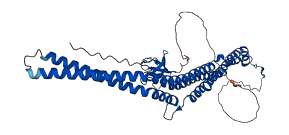
Pacsin1 consists of an N-terminal F-BAR linked to a C-terminal dynamin-binding SH3 domain via a long, flexible tether containing NPF motifs and retains membrane tubulation activity within the F-BAR domain. Pacsin1 F-BAR-mediated membrane deformation is autoinhibited by the SH3 domain. Release from the clamped conformation is driven by binding of Pacsin1 SH3 with the proline-rich domain of dynamin-1.
Autoinhibitory domains (AIDs)
Target domain |
16-273 (The F-BAR, FES-CIP4 Homology and Bin/Amphiphysin/Rvs, domain of Protein kinase C and Casein kinase Substrate in Neurons 1, PACSIN1) |
Relief mechanism |
Partner binding |
Assay |
Deletion assay, Structural analysis |
Accessory elements
No accessory elements
References
- Rao Y et al. (2010) "Molecular basis for SH3 domain regulation of F-BAR-mediated membrane deformation", Proceedings of the National Academy of Sciences of the United States of America, 107, 8213-8
- Senju Y et al. (2011) "Essential role of PACSIN2/syndapin-II in caveolae membrane sculpting", Journal of cell science, 124, 2032-40
- Wang Q et al. (2009) "Molecular mechanism of membrane constriction and tubulation mediated by the F-BAR protein Pacsin/Syndapin", Proceedings of the National Academy of Sciences of the United States of America, 106, 12700-5
Autoinhibited structure
Activated structure

4 structures for Q61644
| Entry ID | Method | Resolution | Chain | Position | Source |
|---|---|---|---|---|---|
| 2X3V | X-ray | 245 A | A/B/C | 1-337 | PDB |
| 2X3W | X-ray | 264 A | PDB | ||
| 2X3X | X-ray | 335 A | PDB | ||
| AF-Q61644-F1 | Predicted | AlphaFoldDB |
4 variants for Q61644
| Variant ID(s) | Position | Change | Description | Diseaes Association | Provenance |
|---|---|---|---|---|---|
| rs33435921 | 352 | T>P | No | Ensembl | |
| rs47514860 | 379 | E>D | No | Ensembl | |
| rs212348917 | 381 | D>E | No | Ensembl | |
| rs235669891 | 382 | A>T | No | Ensembl |
No associated diseases with Q61644
Functions
| Description | ||
|---|---|---|
| EC Number | ||
| Subcellular Localization |
|
|
| PANTHER Family | PTHR23065 | PROLINE-SERINE-THREONINE PHOSPHATASE INTERACTING PROTEIN 1 |
| PANTHER Subfamily | PTHR23065:SF16 | PROTEIN KINASE C AND CASEIN KINASE SUBSTRATE IN NEURONS PROTEIN 1 |
| PANTHER Protein Class |
cytoskeletal protein
actin or actin-binding cytoskeletal protein |
|
| PANTHER Pathway Category |
Huntington disease PACSIN-1 |
|
15 GO annotations of cellular component
| Name | Definition |
|---|---|
| axon terminus | Terminal inflated portion of the axon, containing the specialized apparatus necessary to release neurotransmitters. The axon terminus is considered to be the whole region of thickening and the terminal button is a specialized region of it. |
| COPI-coated vesicle | A vesicle with a coat formed of the COPI coat complex proteins. COPI-coated vesicles are found associated with Golgi membranes at steady state, are involved in Golgi to endoplasmic reticulum (retrograde) vesicle transport, and possibly also in intra-Golgi transport. |
| cytoplasm | The contents of a cell excluding the plasma membrane and nucleus, but including other subcellular structures. |
| cytoplasmic vesicle membrane | The lipid bilayer surrounding a cytoplasmic vesicle. |
| cytosol | The part of the cytoplasm that does not contain organelles but which does contain other particulate matter, such as protein complexes. |
| endosome | A vacuole to which materials ingested by endocytosis are delivered. |
| glutamatergic synapse | A synapse that uses glutamate as a neurotransmitter. |
| myelin sheath | An electrically insulating fatty layer that surrounds the axons of many neurons. It is an outgrowth of glial cells: Schwann cells supply the myelin for peripheral neurons while oligodendrocytes supply it to those of the central nervous system. |
| perinuclear region of cytoplasm | Cytoplasm situated near, or occurring around, the nucleus. |
| photoreceptor ribbon synapse | A ribbon synapse between a retinal photoreceptor cell (rod or cone) and a retinal bipolar cell. These contain a plate-like synaptic ribbon. |
| plasma membrane | The membrane surrounding a cell that separates the cell from its external environment. It consists of a phospholipid bilayer and associated proteins. |
| postsynaptic density membrane | The membrane component of the postsynaptic density. This is the region of the postsynaptic membrane in which the population of neurotransmitter receptors involved in synaptic transmission are concentrated. |
| presynaptic endocytic zone | A specialized region of the plasma membrane and underlying cytoplasm which surround the the active zone, into which synaptic vesicle membranes are recycled following exocytosis. It is especially enriched in endocytic proteins following intense activity. |
| ruffle membrane | The portion of the plasma membrane surrounding a ruffle. |
| synapse | The junction between an axon of one neuron and a dendrite of another neuron, a muscle fiber or a glial cell. As the axon approaches the synapse it enlarges into a specialized structure, the presynaptic terminal bouton, which contains mitochondria and synaptic vesicles. At the tip of the terminal bouton is the presynaptic membrane; facing it, and separated from it by a minute cleft (the synaptic cleft) is a specialized area of membrane on the receiving cell, known as the postsynaptic membrane. In response to the arrival of nerve impulses, the presynaptic terminal bouton secretes molecules of neurotransmitters into the synaptic cleft. These diffuse across the cleft and transmit the signal to the postsynaptic membrane. |
3 GO annotations of molecular function
| Name | Definition |
|---|---|
| cytoskeletal protein binding | Binding to a protein component of a cytoskeleton (actin, microtubule, or intermediate filament cytoskeleton). |
| identical protein binding | Binding to an identical protein or proteins. |
| phospholipid binding | Binding to a phospholipid, a class of lipids containing phosphoric acid as a mono- or diester. |
12 GO annotations of biological process
| Name | Definition |
|---|---|
| actin filament organization | A process that is carried out at the cellular level which results in the assembly, arrangement of constituent parts, or disassembly of cytoskeletal structures comprising actin filaments. Includes processes that control the spatial distribution of actin filaments, such as organizing filaments into meshworks, bundles, or other structures, as by cross-linking. |
| cytoskeleton organization | A process that is carried out at the cellular level which results in the assembly, arrangement of constituent parts, or disassembly of cytoskeletal structures. |
| negative regulation of endocytosis | Any process that stops, prevents, or reduces the frequency, rate or extent of endocytosis. |
| neuron projection morphogenesis | The process in which the anatomical structures of a neuron projection are generated and organized. A neuron projection is any process extending from a neural cell, such as axons or dendrites. |
| plasma membrane tubulation | A membrane tubulation process occurring in a plasma membrane. |
| positive regulation of dendrite development | Any process that activates or increases the frequency, rate or extent of dendrite development. |
| protein localization to membrane | A process in which a protein is transported to, or maintained in, a specific location in a membrane. |
| protein localization to plasma membrane | A process in which a protein is transported to, or maintained in, a specific location in the plasma membrane. |
| regulation of endocytosis | Any process that modulates the frequency, rate or extent of endocytosis. |
| regulation of postsynaptic neurotransmitter receptor internalization | Any process that modulates the frequency, rate or extent of endocytosis of neurotransmitter receptor at the postsynapse. |
| signal transduction | The cellular process in which a signal is conveyed to trigger a change in the activity or state of a cell. Signal transduction begins with reception of a signal (e.g. a ligand binding to a receptor or receptor activation by a stimulus such as light), or for signal transduction in the absence of ligand, signal-withdrawal or the activity of a constitutively active receptor. Signal transduction ends with regulation of a downstream cellular process, e.g. regulation of transcription or regulation of a metabolic process. Signal transduction covers signaling from receptors located on the surface of the cell and signaling via molecules located within the cell. For signaling between cells, signal transduction is restricted to events at and within the receiving cell. |
| synaptic vesicle endocytosis | A vesicle-mediated transport process, in which the synaptic vesicle membrane constituents are retrieved from the presynaptic membrane on the axon terminal after neurotransmitter secretion by exocytosis. Synaptic vesicle endocytosis can occur via clathrin-dependent and clathrin-independent mechanisms. |
15 homologous proteins in AiPD
| UniProt AC | Gene Name | Protein Name | Species | Evidence Code |
|---|---|---|---|---|
| P25623 | SYP1 | Suppressor of yeast profilin deletion | Saccharomyces cerevisiae (strain ATCC 204508 / S288c) (Baker's yeast) | PR |
| A7MBI0 | PACSIN1 | Protein kinase C and casein kinase substrate in neurons protein 1 | Bos taurus (Bovine) | SS |
| O13154 | PACSIN2 | Protein kinase C and casein kinase substrate in neurons protein 2 | Gallus gallus (Chicken) | SS |
| Q9UKS6 | PACSIN3 | Protein kinase C and casein kinase substrate in neurons protein 3 | Homo sapiens (Human) | SS |
| Q9UNF0 | PACSIN2 | Protein kinase C and casein kinase substrate in neurons protein 2 | Homo sapiens (Human) | EV |
| Q9BY11 | PACSIN1 | Protein kinase C and casein kinase substrate in neurons protein 1 | Homo sapiens (Human) | EV |
| Q9H939 | PSTPIP2 | Proline-serine-threonine phosphatase-interacting protein 2 | Homo sapiens (Human) | PR |
| Q99JB8 | Pacsin3 | Protein kinase C and casein kinase II substrate protein 3 | Mus musculus (Mouse) | SS |
| Q9WVE8 | Pacsin2 | Protein kinase C and casein kinase substrate in neurons protein 2 | Mus musculus (Mouse) | EV |
| P97814 | Pstpip1 | Proline-serine-threonine phosphatase-interacting protein 1 | Mus musculus (Mouse) | PR |
| Q99M15 | Pstpip2 | Proline-serine-threonine phosphatase-interacting protein 2 | Mus musculus (Mouse) | PR |
| Q60780 | Gas7 | Growth arrest-specific protein 7 | Mus musculus (Mouse) | PR |
| Q9QY17 | Pacsin2 | Protein kinase C and casein kinase substrate in neurons 2 protein | Rattus norvegicus (Rat) | SS |
| Q9Z0W5 | Pacsin1 | Protein kinase C and casein kinase substrate in neurons protein 1 | Rattus norvegicus (Rat) | SS |
| Q4V920 | pacsin1b | Protein kinase C and casein kinase substrate in neurons protein 1 | Danio rerio (Zebrafish) (Brachydanio rerio) | SS |
| 10 | 20 | 30 | 40 | 50 | 60 |
| MSGSYDEASE | EITDSFWEVG | NYKRTVKRID | DGHRLCNDLM | SCVQERAKIE | KAYAQQLTDW |
| 70 | 80 | 90 | 100 | 110 | 120 |
| AKRWRQLIEK | GPQYGSLERA | WGAMMTEADK | VSELHQEVKN | SLLNEDLEKV | KNWQKDAYHK |
| 130 | 140 | 150 | 160 | 170 | 180 |
| QIMGGFKETK | EAEDGFRKAQ | KPWAKKMKEL | EAAKKAYHLA | CKEERLAMTR | EMNSKTEQSV |
| 190 | 200 | 210 | 220 | 230 | 240 |
| TPEQQKKLVD | KVDKCRQDVQ | KTQEKYEKVL | EDVGKTTPQY | MEGMEQVFEQ | CQQFEEKRLV |
| 250 | 260 | 270 | 280 | 290 | 300 |
| FLKEVLLDIK | RHLNLAENSS | YMHVYRELEQ | AIRGADAQED | LRWFRSTSGP | GMPMNWPQFE |
| 310 | 320 | 330 | 340 | 350 | 360 |
| EWNPDLPHTT | AKKEKQPKKA | EGATLSNATG | AVESTSQAGD | RGSVSSYDRG | QTYATEWSDD |
| 370 | 380 | 390 | 400 | 410 | 420 |
| ESGNPFGGNE | ANGGANPFED | DAKGVRVRAL | YDYDGQEQDE | LSFKAGDELT | KLGEEDEQGW |
| 430 | 440 | ||||
| CRGRLDSGQL | GLYPANYVEA | I |